Posts

Image
After about 3-4 weeks, we have ran 3 trials for each salt concentration. We have one set of data points so far and by Tuesday we will have all 3 trials figured out. The first round of data points all turned out good with no contamination. The process has been good overall and we have not ran into any major issues. In the beginning we had a few hiccups with contamination on our TGY plates but nothing since. We are excited to get the rest of our data points and run the statistics to put a poster together.
Image
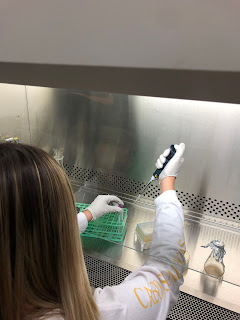
The week of 10/11 and 10/12, Becca and I filled our 96 well plates with 10 micro liters of salt and 90 micro liters of our TGY with Gobi. After this was done, we had to let the bio film grow for 4-5 days. After the growth the next steps were doing a water wash, the CV, and the acetic acid. The process for the water wash and Crystal Violet is to sling, wash with water, pat dry, and repeat 3 times.
Image
On the week of 10/6 we planned out the rest of our experiment that way we were prepared for the process. We figured out the salt concentration and decided on a serial dilution starting with 1M, to 0.1M, 0.01M, 0.001M concentrations. These concentrations are how we will be able to determine the effects of biofilm. The serial dilution started with an eppendorf tube filled with 1000 micro liters of stock salt. The picture below explains the concentrations in each.
Image
On 9/28 we took 3 mL of TGY broth and placed it in a 15 mL flacon tube to prepare for inoculation on Saturday. Chad did a 4 way streak to begin growing the Gobi and allow to inoculate over night. On 9/29 we heated up the TGY broth that has agar to begin plating. The plates had some growth visible but no individual colonies. We did a gram stain on the culture that grow to look for grape like clusters of cells. Rebecca took a cluster of cells straight from the plates into a slide that has 5 micro-liters of TGY broth. The gram stain showed gobi was present but the heat fix caused the cells to turn black instead of purple.
Image
On 9/21 we researched background on biofilm to make sure this is what we wanted to work on. On 9/22 Rebecca and I formed a plan for our research project. We came up with the idea to use different concentrations of several metal ions to test the relation to growth of biofilm. We made a flow chart to map out the process and find an overview of our procedures. So far we have run into the problem of the days not matching with the lab schedules. Although we are off to a slow start, I feel like next week we will really be able to begin our project.

9/14 and 9/15 labs

Image
On 9/14 we did a gram stain on D. Rad. Unfortunately there were not enough cells and we did not heat fix long enough to see any result under the microscope. The cells began to wash away during the process. On 9/15 we decided to reattempt gram staining on P. fluorescens. This time we heat fixed even more so the cells did not wash away. The sample was a bit older coming from August, therefore the cells appeared as debris rather than clumps of cells.
Image
On 9/1 in lab we went over the gram staining process as well as viewed the results under a microscope. There were no complications other than some imperfections on the microscope slide. A large pink oval appeared when viewing. We also observed Chad running his protein gel. These gels are ran vertically rather than the gels we have ran in the past that were done vertically. It took 30 minutes to let the gel bands separate before cracking off the plastic and letting it sit on the rocker over the weekend.  The most important tip Chad gave us was to make sure the gel did not fold on itself.